You must be signed in to read the rest of this article.
Registration on CDEWorld is free. You may also login to CDEWorld with your DentalAegis.com account.
It is estimated that up to 5 million dental implants are placed each year.1 While longitudinal survival rates of osseointegrated dental implants range from 90% to 95%, these numbers represent implants that are present and in function, but may not fully capture rates of peri-implant disease and/or health.2-5 Along with biomechanical complications, implants are susceptible to peri-implant diseases caused by biofilm similar to that seen in periodontal disease.6,7 Peri-implant mucositis is associated with clinical signs of inflammation, in particular bleeding on gentle probing.8 Erythema and swelling may also be present at sites with peri-implant mucositis, but in these cases bone loss has not yet occurred.8 Peri-implantitis is characterized by inflammation in the peri-implant mucosa and subsequent progressive loss of supporting bone.9 Depending on the parameters used to define the disease, estimated rates of peri-implantitis have ranged widely, from 10% to 47% of implants (with a weighted mean prevalence of 22%)10-12 and similarly, rates of peri-implant mucositis have ranged from 19% to 65%, with a weighted mean prevalence of 43%.11 It has also been demonstrated that the risk of peri-implant diseases is increased in patients who smoke and/or have a history of periodontal disease.12-14
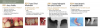
Peri-implant Health
In health, the peri-implant sites are characterized by absence of erythema, bleeding on probing (BOP), swelling, and suppuration. Probing depths are generally deeper at dental implants versus tooth sites and papillae at the interproximal sites of an implant may be shorter than those at interproximal tooth sites, but these have been shown to be compatible with health.15 Histologically, a small number of inflammatory cells may be present in the connective tissue lateral to the sulcular epithelium, coronal to the area of direct bone-to-implant contact.15
Peri-implant Mucositis
Peri-implant mucositis demonstrates clinical signs of inflammation: bleeding upon gentle probing, erythema, and/or swelling.8 An increase in probing depth is often seen when compared to peri-implant health due to swelling of gingival tissues and increased probe penetration into the peri-implant tissues.8 Peri-implant mucositis is strongly associated with dental plaque, and plaque removal has been associated with the resolution of peri-implant mucositis, although clinical and histological resolution of inflammation is significantly slower than at similar gingivitis lesions at teeth.8 Histologically, peri-implant mucositis is characterized by a well-defined inflammatory infiltrate, including plasma cells and lymphocytes with increased vascularity, extending apical to the junctional epithelium into the supracrestal connective tissue zone.8 Data indicate that peri-implant mucositis lesions may progress to peri-implantitis, especially in the absence of regular plaque removal and maintenance.8
Peri-implantitis
Peri-implantitis is a plaque-associated palthological condition characterized by inflammation in the peri-implant mucosa and loss of peri-implant supporting bone.9 Clinically, peri-implantitis is associated with clinical signs of inflammation, BOP, suppuration, increased probing depths, and/or recession of the marginal mucosal tissues. Radiographic bone loss compared to previous examinations is also present.9 Disease progression rates vary and changes in probing depths and radiographic bone levels are useful in determining disease progression rates.9 Histologically, peri-implantitis lesions extend apical to the junctional epithelium and contain high numbers and density of plasma cells, macrophages, and neutrophils.9 Peri-implantitis lesions are generally significantly larger than those at peri-implant mucositis sites.9
Risk Factors for Development of Peri-implant Diseases
In clinical practice, successful dental implant therapy allows practitioners to help patients achieve optimal oral health and esthetic outcomes. These outcomes rely heavily upon many patient, site, and treatment-related factors.8,9,16 Factors related to patients' systemic health, behaviors, site-specific anatomy, restoration type, and fixture type have all been indicated in the development of peri-implant diseases and worsening treatment outcomes after therapy.8,9,16 Proper patient selection and pre-procedural planning to reduce these risk factors may reduce the overall need for peri-implant disease treatment. Specific risk factors are discussed below. For the purposes of risk stratification and discussion of risk mitigation, risk factors are identified as biologic or prosthetic/occlusal (Figure 1).
Biologic Risk Factors
1. Systemic Diseases
Dental implants are generally elective surgical procedures and should be undertaken on patients who are systemically healthy enough to undergo elective, outpatient procedures. Some common systemic diseases may also directly affect the rates of implant survival. While data are equivocal with regard to the role of glycemic control in implant failure rates, dental implants placed in patients with diabetes have been shown to have higher failure rates than those placed in nondiabetic patients.17,18 This is generally believed to be associated with levels of glycemic control and the biologic complications of hyperglycemia.19 In humans, hyperglycemia is known to impair wound healing, host defense against pathogens, and new bone formation and bone repair, while prolonging the inflammatory response to injury.18 The recommended osseointegration periods may be extended in diabetics, based on level of glycemic control, due to the delay in wound healing caused by hyperglycemia.20 Future studies are needed to identify distinct cut-off points, fully assess the risk conveyed by the duration of disease, and quantify the risks, if any, associated with hyperglycemia levels and development of peri-implantitis.
Osteoporosis may also potentially affect implant survival. Osteoporosis and osteopenia are diseases characterized by low bone mass and micro-architectural deterioration with a consequent increase in bone fragility and susceptibility to fracture.21,22 While osteoporosis/osteopenia have common risk factors with periodontal and peri-implant diseases, including cigarette smoking, dietary factors, and medications, periodontal disease has been independently associated with osteoporotic status.23 Peri-implantitis, periodontal disease, and osteoporosis are mediated by similar dysfunction in the bone remodeling process and the interaction between these diseases may be expected.23 Patients with osteoporosis demonstrated decreased alveolar and axial bone density and mass and thinner cortical bone than healthy counterparts.24 To date, studies have not shown a definitive association of peri-implantitis with osteoporosis or osteopenia, although implant placement and use of bisphosphonate medications have been shown to potentially mitigate alveolar bone loss in osteoporotic patients.9,16,25-27
2. Medications
Medications that act centrally or locally to alter healing, bone metabolism, immune response, and/or cell-to-cell interactions may have either positive or negative effects on implant healing and survival.28 Both selective serotonin reuptake inhibitors (SSRIs) and proton pump inhibitors (PPIs) have been demonstrated to be associated with decreased osseointegration and implant survival.29-31 Conversely, beta-blocker antihypertensives, statin medications, and oral bisphosphonate medications have been shown to be associated with improved outcomes of dental implants when compared to individuals with similar medical conditions taking placebo.32-34 Perioperative, short-term non-steroidal anti-inflammatory drug (NSAID) use has been associated in some studies with increased risk of implant failure, but this is contradicted by other research.35,36 In light of emerging evidence of the influence of systemic medications on short- and long-term implant success, a thorough medication review and critical informed consent discussion, including the risks/benefits posed by medications for other conditions, must be performed by dental healthcare providers as a part of the pre-operative evaluation.
3. Smoking Status and/or Tobacco Cessation
Smoking has been shown to have many negative effects on the oral cavity and wound healing after procedures, such as reduction in neutrophil chemotactic response, vasoconstriction, alterations in immune response, an increase in number or proportion of periopathogenic bacteria, and a decrease in fibroblast number and collagen production.9,37 These effects of smoking can lead to chronic inflammation at periodontal and peri-implant tissues. Patients who smoke have been shown to have up to two times the failure rate of implants compared to non-smokers and that smoking itself is a predisposing factor for peri-implantitis.9 Smoking cessation is an important contributing factor to implant success; even though cessation cannot reverse past effects, it can increase implant success rates to that of nonsmokers.9 In a large epidemiologic trial, each additional year of smoking cessation was associated with a 3.9% reduction in risk for periodontal attachment loss,38 but the influence on peri-implantitis is murkier, likely due to confounding with a history of periodontitis. In patients who use tobacco, maintenance is increasingly critical as supportive implant therapy for smokers shows a greater benefit in reducing rates of peri-implantitis than in non-smokers.39
4. Periodontal Health
A history of periodontitis is a risk factor for peri-implantitis.5,8,9,15,40 The two diseases share a common primary etiology of anaerobic bacteria.41 Findings suggest that bacteria associated with periodontal disease and peri-implant diseases are similar.41,42 The principal pathogens in peri-implant disease are P. gingivalis and A. actinomycetemcomitans, known keystone pathogens in periodontitis.42 Colonization with these bacterial species occurs within the first 28 days after implant exposure to the oral environment,43 and bacteria can be transferred from distant reservoirs, such as deep probing depths at teeth elsewhere within a patient's mouth.44 As periodontitis is the reason for the majority of tooth loss in adult patients, treatment of active periodontitis and continued maintenance therapy are essential to success in patients who will then have tooth replacement with dental implant therapy.40
5. Plaque Control and Adherence to Regular Supportive Implant Therapy
Plaque control performed by patients and dental professionals can result in a reduction in clinical signs of peri-implant inflammation.45 Bacterial load and pathogenicity of oral biofilm have been associated with increased development of peri-implant diseases.45 Therefore, regular maintenance protocols that reduce overall bacterial loads and alter the qualitative bacterial make-up are critical to reduce bacteria at teeth and implants and decrease the likelihood of transmission of periodontal pathogenic bacteria to implant sites.46 Additionally, lack of adherence to supportive peri-implant therapy results in significantly higher frequencies of sites with mucosal inflammation and peri-implant bone loss.45 Tailored peri-implant maintenance strategies, such as personalized oral hygiene techniques, implant examination and imaging, and professional implant cleaning must be an ongoing component of all implant treatment plans.45
6. Peri-implant Soft Tissue Quality/Quantity
While the importance of keratinized and/or attached mucosa around teeth and implants is equivocal for their survival, it has been proposed that the establishment of a circumferential seal of tightly packed collagen around the implant-oral cavity interface may improve long-term implant success.2 Survival rates have been shown to be equivalent for implants placed in keratinized and alveolar mucosa, but increased radiographic bone loss and higher levels of gingival inflammation are associated with a lack of keratinized mucosa.47-49 While there are no definitive studies to conclude that there is a benefit when implants have an adequate band of fixed and/or keratinized mucosa, in patients with other risk factors, including increased plaque accumulation and previous history of periodontitis, increased keratinized mucosa may be protective to allow for personal and professional plaque removal.47-49
Prosthetic/Occlusal Risk Factors
1. Prosthetic Design and Occlusal Load
It has been postulated that mechanical overloading is one of the main reasons for peri-implant bone loss and late implant failures.3 Occlusal load is influenced by prosthetic design, but hard to study due to lack of quantification of overload. Interfacial micromotion compromises the establishment of implant osseointegration during early healing.50 Additionally, implants demonstrating off-axis forces exhibit more peri-implant bone loss after loading.41 Despite the elusive nature of occlusal overload quantification, a systematic review concluded that occlusal overloading was associated with peri-implant marginal bone loss caused by microtrauma concentrated at the marginal bone.51 It follows that prosthetically-driven treatment planning as well as assessment of occlusal load, including periodic inspection of implant prostheses for signs of wear and parafunctional habits, and occlusal adjustment when premature contacts or interferences are present, should be undertaken during the maintenance phase.4
2. Retained Cement
Retained cement has been indicated in a large number of peri-implant disease cases.8,52 Many dental implant cements are radiolucent or poorly radiopaque, and residual cement may not be detected radiographically, particularly if present on the buccal and/or lingual of the fixture. Residual cement presents a rough surface and may allow bacterial attachment, subsequently leading to peri-implant inflammation.53 Peri-implant disease prevalence is significantly higher at fixtures with cement-retained versus screw-retained restorations.52,53 Additionally, in a case-control study of implants with peri-implantitis, 81% had excess cement present, whereas no retained cement was found at healthy, control implants.52 To reduce the risk of retention of excess cement, practitioners are cautioned to practice techniques such as use of screw-retained restorations, allowing for adequate soft tissue healing prior to seating of permanent restoration, use of extraoral implant/abutment analogs to reduce residual cement prior to insertion, and early follow-up after initial cementation to detect any early signs of cement retention.
3. History of Parafunctional Habits/Occlusal Dysfunction
There are fundamental differences between teeth and implants as they relate to biomechanics and attachment apparatus. Dental implants lack a periodontal ligament and, instead, exhibit osseointegration, or functional ankylosis.2,54 This attachment allows for considerably less axial movement in response to occlusal forces than seen in teeth, 3-5 μm and 25-100 μm,55,56 respectively. This functional difference also results in stress concentration at the crestal bone of implants as compared with stress distribution and shock absorption in teeth.55 Because of this, occlusal overload at dental implants can result in significant prosthetic and osseointegration failure, including abutment screw loosening or fracture, abutment or prosthesis fracture, implant fracture, and/or bone loss.57 While off-axis forces have not been shown to decrease implant and prosthesis survival in cases with cross-arch stabilization,58-61 prosthetic complications and marginal bone loss may be greater, particularly in cases with implant angulation > 30°.57,62 Assessment of occlusal patterns and parafunctional habits as well as implant placement and prosthetic design minimizing large cantilevers, off-axis and excessive force, overly broad occlusal table size, and parafunction should be undertaken to reduce prosthetic and implant complications. Additionally, it has been reported that there is insufficient data in the current literature to establish firm clinical guidelines for implant occlusion, and there is a need for further studies in the future.57,63
Diagnosis of Peri-Implant Diseases
Diagnosis of peri-implant conditions requires good baseline assessment and frequent monitoring of implants with clinical and radiographic examination.8,9 Radiographs that clearly show implant threads to ensure that they are parallel should be taken at placement and final prosthetic seating. A clinical examination at the time of final prosthetic seating should be performed to establish baseline probing depths and ensure that BOP and suppuration are not present. It is also recommended that radiographs be taken to assess bone levels at regular intervals during implant healing and maintenance. A simplified diagnostic rubric should be applied to implant assessment at each maintenance visit (Figure 2). The protocol is briefly described below.
Step 1: Is mobility present?
• Mobility should be assessed and the source of the mobility determined. Moblity may be due to abutment loosening, abutment screw fracture, prosthesis fracture, failure/loss of osseointegration, or implant fracture.
Step 2: Are the clinical signs of inflammation present?
• The presence of BOP and/or suppuration should be assessed. If BOP or suppuration is present, peri-implant health is not a possible diagnosis and further investigation should be performed to distinguish between peri-implant mucositis and peri-implantitis.
• Probing depths should be taken at 4-6 sites per implant and compared with baseline values.
Step 3: Are there changes in interproximal bone levels from the baseline evaluation?
• Standard periapical radiographs taken parallel to the implant body so that the threads are distinct will allow evaluation of the interproximal bone levels. If bone levels differ at the mesial and distal aspect, the areas with the greatest bone loss should be used in the assessment. Bone loss greater than that associated with initial remodeling, which may be specific to the implant design and surface topography, in addition to clinical signs of inflammation, is considered diagnostic for peri-implantitis.
• Cone beam computerized tomography may also be used to assess bone levels in 3 dimensions, including that at the direct buccal and lingual of the dental implant, but in most cases periapical radiographs are adequate to make diagnosis. Understanding pixel size and diagnostic capabilities of the particular radiographic modality, including scatter from metal-base restorations. Caution should be taken by practitioners to adhere to ALARA principles and reduce patietnts' radiographic exposure to the lowest level that allows for adequate diagnostic abilities.
Step 4: Are there any iatrogenic or mutable factors that may contribute to inflammation, plaque retention, and /or bone loss?
• Retained cement, inadequate restorative seating/misfit, open contacts, occlusal overload, and/or prosthesis overcontour should be considered and addressed to allow for adequate plaque control.
Implant Maintenance Protocols
In multiple studies, poor adherence to prescribed implant maintenance was associated with higher levels of peri-implant mucosal inflammation and peri-implant bone loss as well as more frequent implant loss.44,45,64,65 Regular implant maintenance has also been demonstrated to be the most cost-effective intervention for long-term implant success.39 It is well-established that periodontal maintenance improves the health and retention of natural teeth.66,67 When contrasting teeth and implants, human and animal studies have demonstrated more rapid progression of inflammatory lesions after plaque accumulation and slower resolution of the lesion at implant versus tooth sites.8,9 Given these similarities, it follows that implant maintenance improves long-term implant outcomes and should be based upon individual patient risk stratification and diagnoses.44,45,64,65 In everyday practice, this means that ongoing monitoring, diagnosis of peri-implant conditions, utilizing patient-centered motivation to improve plaque control, professional peri-implant plaque removal, and control of modifiable risk factors.
Professional Treatment of Peri-implant Diseases
Peri-implant mucositis has been shown to be reversible with nonsurgical therapy and improved plaque control, but gold-standard treatment for peri-implantitis lesions remains elusive.5,8,9,41,42 Nonsurgical therapy may reduce clinical signs of inflammation in the short-term at peri-implantitis lesions, but it has been demonstrated to have limited effectiveness for long-term therapy.8,9,68,69 Nevertheless, mechanical removal of oral biofilm and calculus should be undertaken to remove inflammation and improve outcomes of other interventions.8,9,68,69 Manual instruments should be limited to metals softer than or of the same hardness as the titanium. Graphite, gold-platinum alloy, or titanium curettes are generally used for this purpose.68,70 Additionally, the use of implant-specific ultrasonic inserts/tips may be beneficial in providing increased lavage and mechanical biofilm removal.68,70 Surface alteration of titanium, type II gold, and porcelain dental materials is significantly less with either copper alloy or carbon composite ultrasonic tips when compared to conventional metal. Zirconia materials have not shown such increased roughness when treated with conventional metal instruments.71 Chemotherapeutic agents are considered an integral part of implant surface decontamination for titanium implants with peri-implantitis, but some professionally used chemotherapeutic agents including chlorhexidine have also been shown to compromise the biocompatibility of titanium.72,73 Sterile saline, citric acid, and EDTA have been proposed to demonstrate adequate biocompatibility and may be used for professional implant surface decontamination.72,73 Surgical intervention may be undertaken to reduce deep peri-implant probing depths and/or repair peri-implant intrabony defects.69,70The efficacy of these approaches may vary based upon the extent, chronicity, and underlying etiology of the lesion and the surgical techniques employed.68-70
Summary
Dental implants are a common and successful method of tooth replacement. However, the prevalence of peri-implant diseases is high and dental healthcare professionals must be aware of underlying risk for peri-implant diseases during implant treatment planning, as well as the importance of a customized implant maintenance protocol should be stressed.
Clinical Recommendations
• Proper assessment of the patient to identify risk factors for peri-implant disease should be undertaken prior to implant placement.
• Modification of any mutable risk factors, including treatment of active periodontal disease, should be completed prior to implant therapy.
• Baseline clinical evaluation and radiographs should be established as a comparison for future implant assessments.
• Individualized maintenance therapy should be developed for each patient, taking into account risk factors, compliance, and oral hygiene.
• Surveillance of implants to identify early signs of peri-implant disease should be done on frequent individualized intervals.
• Effective removal of oral plaque biofilm, along with patient motivation to ensure optimal oral hygiene and compliance with maintenance, is a critical part of therapy to improve implant outcomes.
About the Author
Maria L. Geisinger, DDS, MS
Associate Professor
University of Alabama at Birmingham
Department of Periodontology
References
1. Johnson J. Dental implants. ADA Patient Smart Patient Education Center. http://www.ada.org/~/media/ADA/Publications/Files/ADA_PatientSmart_Implants.ashx. Published 2014. Accessed May 15, 2019.
2. Brånemark PI, Hansson BO, Adell R, et al. Osseointegrated implants in the treatment of the endentulous jaw. Experience from a 10-year period. Scand J Plast Reconstr Surg Suppl.1977;16:1-132.
3.Esposito M, Hirsch JM, Lekholm U, Thomsen P. Biological factors contributing to failures of osseointegrated oral implants. (II). Etiopathogenesis. Eur J Oral Sci.1998;106(3):721-764.
4. Ashey ET, Covington LL, Bishop BG, Breault LG. Ailing and failing endosseous dental implants: a literature review. J Contemp Dent Pract.2003;4(2):35-50.
5. Rosen P, Clem D, Cochran DL, et al; American Academy of Periodontology Task Force on Peri-Implantitis. Academy report: peri-implant mucositis and peri-implantitis: a current understanding of their diagnoses and clinical implications. J Periodontol.2013;84(4):436-443.
6. Jepsen S, Berglundh T, Genco R, et al. Primary prevention of peri-implantitis: managing peri-implant mucositis. J Clin Periodontol.2015;42(suppl 16):S152-S157.
7. Berglundh T, Armitage G, Araujo MG, et al. Peri-implant diseases and conditions: consensus report of workgroup 4 of the 2017 World Workshop on the Classification of Periodontal and Peri-implant Diseases and Conditions. J Periodontol.2018;89(suppl 1):S313-S318.
8. Heitz-Mayfield LJA, Salvi GE. Peri-implant mucositis. J Periodontol.2018;89(suppl 1):S257-S266.
9. Schwarz F, Derks J, Monje A, Wang HL. Peri-implantitis. J Periodontol.2018;89(suppl 1):S267-S290.
10. Mombelli A, Müller N, Cionca N. The epidemiology of peri-implantitis. Clin Oral Implants Res.2012;23(suppl 6):67-76.
11. Derks J, Tomasi C. Peri-implant health and disease: a systematic review of current epidemiology. J Clin Periodontol.2015;42(suppl 16):S158-S171.
12. Fransson C, Wennström J, Tomasi C, Berglundh T. Extent of peri-implantitis-associated bone loss. J Clin Periodontol.2009;36(4):357-363.
13. Atieh MA, Alsabeeha NH, Faggion CM Jr. Duncan WJ. The frequency of peri-implant diseases: a systematic review and meta-analysis. J Periodontol.2013;84(11):1586-1598.
14. Sgolastra F, Petrucci A, Severina M, et al. Periodontitis, implant loss and peri-implantitis: a meta-analysis. Clin Oral Implants Res.2015;26(4):e8-e16.
15. Araujo MG, Lindhe J. Peri-implant health. J Periodontol.2018;89(suppl 1):S249-S256.
16. Curtis DA, Lin GH, Fishman A, et al. Patient-centered risk assessment in implant treatment planning. Int J Oral Maxillofac Implants.2019;34(2):506-520.
17. Morris HF, Ochi S, Winkler S. Implant survival in patients with type 2 diabetes: placement to 36 months. Ann Periodontol.2000;5(1):157-165.
18. Iacopino AM. Periodontitis and diabetes interrelationships: role of inflammation. Ann Periodontol.2001;6(1):125-137.
19. Naujokat H, Kunzendorf B, Wiltfang J. Dental implants and diabetes mellitus-a systematic review. Int J Implant Dent. 2016;2(1):5.
20. Oates TW, Huynh-Ba G, Vargas A, et al. A critical review of diabetes, glycemic control, and dental implant therapy. Clin Oral Implants Res. 2013;24(2):117-127.
21. Kanis JA. Assessment of fracture risk and its application to screening for postmenopausal osteoporosis: synopsis of a WHO report. WHO Study Group. Osteoporos Int. 1994;4(6):368-381.
22. Kanis JA, Melton LJ III, Christiansen C, et al. The diagnosis of osteoporosis. J Bone Miner Res. 1994;9(8):1137-1141.
23. Geurs NC. Osteoporosis and periodontal disease. Periodontol 2000. 2007;44:29-43.
24. Wehren LE. The epidemiology of osteoporosis and fractures in geriatric medicine. Clin Geriatr Med.2003;19(2):245-258.
25. Ruggiero SL, Mehrotra B, Rosenberg TJ, Engroff SL. Osteonecrosis of the jaws associated with the use of bisphosphonates: a review of 63 cases. Int J Oral Maxillofac Surg. 2004;62(5):527-534.
26. Kribbs PJ, Smith DE, Chesnut CH III. Oral findings in osteoporosis. Part I: measurement of mandibular bone density. J Prosthet Dent. 1983;50(4):576-579.
27. Geurs NC, Lewis CE, Jeffcoat MK. Osteoporosis and periodontal disease progression. Periodontol 2000. 2003;32:105-110.
28.Ibraheem A, Batra C, John V, Shin D. Role of medication in osseointegration of dental implants. Decisions in Dentistry.2019;5(5):14-21.
29. Wu X, Al-Abedalla K, Rastikerdar E, et al. Selective serotonin reuptake inhibitors and the risk of osseointegrated implant failure: a cohort study. J Dent Res.2014;93(11):1054-1061.
30. Wu X, Al-Abedalla K, Abi-Nader S, et al. Proton pump inhibitors and the risk of osseointegrated dental implant failure: a cohort study. Clin Implant Dent Relat Res.2017;19(2):222-232.
31. Chappius V, Avila-Ortiz G, Araújo MG, Monje A. Medication-related dental implant failure: systematic review and meta-analysis. Clin Oral Implants Res.2018;29(suppl 16):55-68.
32. Al-Subaie AE, Laurenti M, Abdallah MN, et al. Propranolol enhances bone healing and implant osseointegration in rats tibiae. J Clin Periodontol.2016;43(12):1160-1170.
33. Gelazius R, Poskevicius L, Sakavicius D, et al. Dental implant placement in patients on bisphosphonate therapy: a systematic review. J Oral Maxillofac Res.2018;9(3):e2.
34. Du Z, Chen J, Yan F, Xiao Y. Effects of simvastatin on bone healing around titanium implants in osteoporotic rats. Clin Oral Implants Res.2009;20(2):145-150.
35. Winnett B, Tenenbaum HC, Ganss B, Jokstad A. Perioperative use of non-steroidal anti-inflammatory drugs might impair dental implant osseointegration. Clin Oral Implants Res.2016;27(2):e1-7.
36. Fu JH, Bashutski JD, Al-Hezaimi K, Wang HL. Statins, glucocorticoids, and nonsteroidal anti-inflammatory drugs: their influence on implant healing. Implant Dent.2012;21(5):362-367.
37. Schwarz F, Becker K, Sahm N, et al. The prevalence of peri-implant diseases for two-piece implants with an internal tube-in-tube connection: a cross-sectional analysis of 512 implants. Clin Oral Implants Res.2017;28(1):24-28.
38. ALHarthi SSY, Natto ZS, Midle JB, et al. Association between time since quitting smoking and periodontitis in former smokers in the National Health and Nutrition Examination Surveys (NHANES) 2009 to 2012. J Periodontol.2019;90(1):16-25.
39. Schwendicke F, Tu YK, Stolpe M. Preventing and treating peri-implantitis: a cost-effectiveness analysis. J Periodontol.2015;86(9):1020-1029.
40. Roccuzzo M, De Angelis N, Bonino L, Aglietta M. Ten-year results of a three-arm prospective cohort study on implants in periodontally compromised patients. Part 1: implant loss and radiographic bone loss. Clin Oral Implants Res. 2010;21(5):490-496.
41. Mombelli A, Lang NP. The diagnosis and treatment of peri-implantitis. Periodontol 2000. 1998;17:63-76.
42. Ata-Ali J, Candel-Marti ME, Flichy-Fernández AJ, et al. Peri-implantitis: associated microbiota and treatment. Med Oral Patol Oral Cir Bucal. 2011;16(7):e937-e943.
43. Renvert S, Roos-Jansåker AM, Lindahl C, et al. Infection at titanium implants with or without a clinical diagnosis of inflammation. Clin Oral Implants Res. 2007;18(4):509-516.
44. Quirynen M, Vogels R, Peeters W, et al. Dynamics of subgingival colonization of ‘pristine' peri-implant pockets. Clin Oral Implants Res. 2006;17(1):25-37.
45. Ramanauskaite A, Tervonen T. The efficacy of supportive peri-implant therapies in preventing peri-implantitis and implant loss: a systematic review of the literature. J Oral Maxillofac Res.2016;7(3):e12.
46. Gouvoussis J, Sindhusake D, Yeung S. Cross-infection from periodontitis sites to failing implant sites in the same mouth. Int J Oral Maxillofac Implants. 1997;12(5):666-673.
47. Souza AB, Tormena M, Matarazzo F, Araújo MG. The influence of peri-implant keratinized mucosa on brushing discomfort and peri-impaltn tissue health. Clin Oral Implants Res.2016;27(6):650-655.
48. Wennström JL, Derks J. Is there a need for keratinized mucosa around implants to maintain health and tissue stability? Clin Oral Implants Res.2012;23(suppl 6):136-146.
49. Lin GH, Chan HL, Wang HL. The significance of keratinized mucosa on implant health: a systematic review. J Periodontol.2013;84(12):1755-1767.
50. Pesce P, Canullo L, Grusovin MG, et al. Systematic review of some prosthetic risk factors for periimplantitis. J Prosthet Dent.2015;114(3):346-350.
51. Fu JH, Hsu YT, Wang HL. Identifying occlusal overload and how to deal with it to avoid marginal bone loss around implants. Eur J Oral Implantol. 2012;5(suppl):S91-S103.
52. Linkevicius T, Puisys A, Vindasiute E, et al. Does residual cement around implant-supported restorations cause peri-implant disease? A retrospective case analysis. Clin Oral Implants Res.2013;24(11):1179-1184.
53. Staubli N, Walter C, Schmidt JC, et al. Excess cement and the risk of peri-implant disease - a systematic review. Clin Oral Implants Res. 2017;28(10):1278-1290.
54. Schroeder A, Polher O, Sutter F. Tissue reaction to an implant of a titanium hollow cylinder with a titanium surface spray layer [in German]. SSO Schweiz Monatsschr Zahnheilkd. 1976;86(7):713-727.
55. Sekine H, Komiyama Y, Potta H, Yoshida K. Mobility characteristics and tactile sensitivity of osseointegrated fixture-supporting systems. In: van Steenberghe D, Albrektsson T, Branemark PI, eds. Tissue Integration in Oral Maxillofacial Reconstruction. Amsterdam: Excerpta Medica; 1986:326-332.
56. Schulte W. Implants and the periodontium. Int Dent J. 1995;45(1):16-26.
57. Kim Y, Oh TJ, Misch CE, Wang HL. Occlusal considerations in implant therapy: clinical guidelines with biomechanical rationale. Clin Oral Implants Res.2005;16(1):26-35.
58. Babbush CA, Kutsko GT, Brokloff J. The all-on-four immediate function treatment concept with NobelActive implants: a retrospective study. J Oral Implantol. 2011;37(4):431-445.
59. Crespi R, Vinci R, Capparé P, et al. A clinical study of edentulous patients rehabilitated according to the "all on four" immediate function protocol. Int J Oral Maxillofac Implants. 2012;27(2):428-434.
60. Ata-Ali J, Peñarrocha-Oltra D, Candel-Marti E, Peñarrocha-Diago M. Oral rehabilitation with tilted dental implants: a metaanalysis. Med Oral Patol Oral Cir Bucal. 2012;17(4):e582-e587.
61. Del Fabbro M, Ceresoli V. The fate of marginal bone around axial vs. tilted implants: a systematic review. Eur J Oral Implantol.2014;7(suppl 2):S171-S189.
62. Hsu ML, Chen FC, Kao HC, Cheng CK. Influence of off-axis loading of an anterior maxillary implant: a 3-dimensional finite element analysis. Int J Oral Maxillofac Implants.2007;22(2):301-309.
63. Koyano K, Esaki D. Occlusion on oral implants: current clinical guidelines. J Oral Rehabil.2015;42(2):153-161.
64. Schwarz F, Becker K, Sager M. Efficacy of professionally administered plaque removal with or without adjunctive measures for the treatment of peri-implant mucositis. A systematic review and meta-anlysis. J Clin Periodontol.2015;42(suppl 16):S202-S213.
65. Monje A, Aranda L, Diaz KT, et al. Impact of maintenance therapy for the prevention of peri-impant diseases: a systematic review and meta-analysis. J Dent Res.2016;95(4):372-379.
66. Axelsson P, Nyström B, Lindhe J. The long-term effect of a plaque control program on tooth mortality, caries, and periodontal disease in adults. Results after 30 years of maintenance. J Clin Periodontol.2004;31(9):749-757.
67. Armitage GC, Xenoudi P. Post-treatment supportive care for the natural dentition and dental implants. Periodontol 2000.2016;71(1):164-184.
68. Lang NP, Salvi GE, Sculean A. Nonsurgical therapy for teeth and implants-When and why? Periodontol 2000.2019;79(1):15-21.
69. Figuero E, Graziani F, Sanz I, et al. Management of peri-implant mucositis and peri-implantitis. Periodontol 2000.2014;66(1):255-273.
70. Romanos GE, Javed F, Delgado-Ruiz RA, Calvo-Guirado JL. Peri-implant diseases: a review of treatment interventions. Dent Clin North Am.2015;59(1):157-178.
71. Seol HW, Heo SJ, Koak JY, et al. Surface alterations of several dental materials by a novel ultrasonic scaler tip. Int J Oral Maxillofac Implants.2012;27(4):801-810.
72. Kotsakis GA, Lan C, Barbosa J, et al. Antimicrobial agents used in the treatment of peri-implantitis alter the physiochemistry and cytocompatibility of titanium surfaces. J Periodontol.2016;87(7):809-819.
73. Subramani K, Wismeijer D. Decontamination of titanium implant surface and re-osseointegration to treat peri-implantitis: a literature review. Int J Oral Maxillofac Implants.2012;27(5):1043-1054.